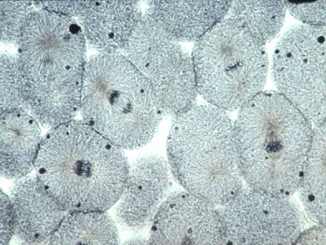
Whitefish Mitosis

A section of whitefish blastula at 400x
Introduction
Why are whitefish blastula used to study mitosis? The blastula is an early stage of embryo development and represents a period in the organism's life when most of the cells are constantly dividing. Moreover, the dividing cell have very large and easily seen chromosomes, so its easy to find lots of cells in each stage of mitosis.Human chromosomes on the other hand, are are not clearly visible at higher power magnification. So, for student purposes, whitefish blastula are used.
Interphase

Whitefish blastula cells in interphase
The three phases of Interphase
G1 Phase
Growth: the cell grows in size and carry out their normal day to day activities.
S Phase
Prior to mitosis, the cell readies itself by duplicating its chromosomes and other cellular contents. The chromosomes at this stage are dispersed and not visible using a light microscope. Before DNA synthesis, each of the cell’s chromosomes consist of one chromatid. After DNA synthesis, each of the cell’s chromosomes consist of 2 genetically identical sister chromatids attached at the centromere.
G2 Phase
The cell prepares the enzymes and machinery for mitosis.
Prophase


Whitefish blastula cells in early prophase
During early prophase, the chromosomes condense, making them distinguishable when using a light microscope.
The nuclear envelope disperses.During late prophase, the nucleoli disappear and the mitotic spindle apparatus assembles.The mitotic spindle will consist of microtubules that extend from pole to pole.
Metaphase


Whitefish blastula cells in metaphase: the cell on the left is in early metaphase, the cell on the right in late metaphase
The mitotic spindle has attached to the centromere of each chromosome and moves them through the "dance of mitosis". Note that the centromeres of each chromosome are aligned at the equator of the cell and the telomeres (ends of the chromosomes) drift away from the equator.
Anaphase

A whitefish blastula cell in anaphase
During anaphase the mitotic spindle apparatus pulls the sister chromatids of each chromosome apart by attaching to each centromere and then pull the chromatids to each pole of the cell. Note that the telomeres of each chromosome point toward the cell’s equator.
Telophase

A whitefish blastula cell in telophase + cytokinesis
Chromosomes begin to disperse. Spindle fibers disperse. Cytokinesis begins--formation of daughter cells. In animals, like the whitefish, a cleavage furrow, a contractile ring of muscle like fibers, pinches the cell into two. The nuclear envelope forms again around the nuclei.
Whitefish Mitosis Practice
|
The photomicrographs below show sections of whitefish blastula. The blastula is an early stage of embryo development, and represents a period in the organism's life when most of the cells are dividing consistently. Practice locating each of the stages of mitosis in the following photomicrographs. Each picture contains at least one cell at each stage of mitosis (and some stages are represented by multiple cells). |